
Blog
1. Title:
Automatic detection and segmentation of morphological changes of the maxillary sinus mucosa on cone-beam computed tomography images using a three-dimensional convolutional neural network
2. Authors: Kuo Feng Hung; Qi Yong H. Ai; Ann D. King; Michael M. Bornstein; Lun M. Wong; Yiu Yan Leung;
3. Summary:
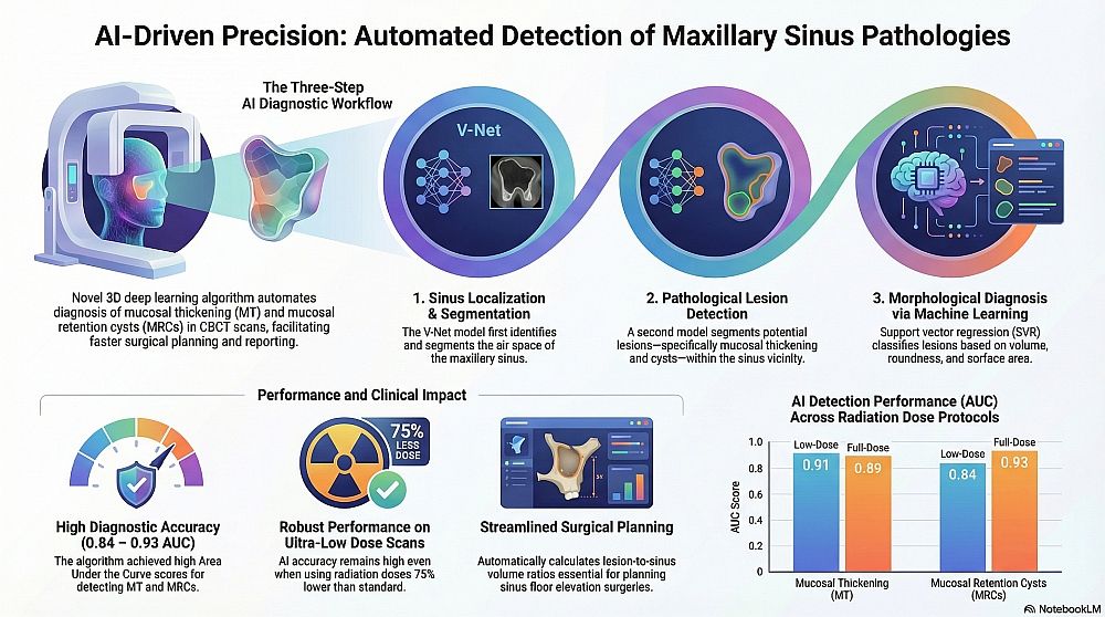
This study proposes and evaluates a three-step artificial intelligence algorithm combining a 3D Convolutional Neural Network (V-Net) and Support Vector Regression (SVR) for the automatic detection and segmentation of mucosal thickening (MT) and mucosal retention cysts (MRCs) in the maxillary sinus. The study utilized 445 Cone-Beam Computed Tomography (CBCT) scans (890 sinuses), divided into low-dose (used for training and testing) and full-dose (used for testing only) datasets. The algorithm demonstrated high diagnostic accuracy (AUC > 0.84) and favorable segmentation performance (DSC > 0.66) on both low-dose and full-dose scans, suggesting its potential for automated diagnosis and treatment planning in daily clinical practice.
4. Key Words: Maxillary sinus; Mucosal thickening; Mucosal retention cyst; Artificial Intelligence; Convolutional neural network; Cone-beam computed tomography
5. Extracted Data
5.1. Year: 2022
5.2. Modality: Cone-Beam Computed Tomography (CBCT) – low-dose and full-dose
5.3. Dataset: 445 CBCT scans (890 maxillary sinuses), consisting of 424 low-dose scans and 21 full-dose scans
5.4. Dataset Split:
5.5. Network Architecture:
##1. A 3D CNN (V-Net) for maxillary sinus air space segmentation.
##2. A second V-Net for segmenting lesions (MT and MRCs).
##3. Support Vector Regression (SVR) for diagnosis/classification based on shape features
5.6. Metrics:
5.7. AP - Professional Qty: 2 annotators
5.8. AP - Supervisor Presence: No info (consensus )
5.9. AP - Experience Level: >6 years
5.10. AP - Expertise Area: OMFR 5.11. AP - Tool or System: ITK-SNAP 3.8.0
5.12. ML Task: 3D Semantic Segmentation; Lesion detection; Morphological Classification
5.13. Project Objective:
To develop and validate a fully automatic system capable of detecting, segmenting, differentiating, and volumetrically quantifying MT and MRC in CBCT images.
6. Clinical Relevance:
The algorithm has the potential to be implemented in daily practice as an automated diagnostic and reporting system. It can visualize pathological findings, measure volumes, and assist in treatment planning for sinus-related oral surgeries, particularly sinus floor elevation (SFE), by alerting clinicians to the presence and extent of sinus opacification