Deep Learning for Alveolar Bone Loss Measurement in Periapical Radiographs
Title: Use of the deep learning approach to measure alveolar bone level
Authors: Chun-Teh Lee; Tanjida Kabir; Jiman Nelson; Sally Sheng; Hsiu-Wan Meng; Thomas E. Van Dyke; Muhammad F. Walji; Xiaoqian Jiang; Shayan Shams
Journal: Journal of Clinical Periodontology
Year: 2022
DOI: https://doi.org/10.1111/jcpe.13574
Summary (Dental AI)
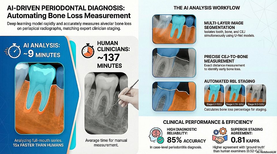
Summary: This study developed a deep learning (DL) model to aid in periodontal diagnosis by measuring radiographic bone level (RBL) using periapical radiographs. The model integrates three segmentation networks (U-Net based) to segment the bone area, teeth, and cemento-enamel junction (CEJ). It calculates RBL percentage and assigns RBL stages and periodontal diagnosis based on the 2018 periodontitis classification. The model demonstrated high accuracy (DSC > 0.91) and reliability comparable to independent examiners, while being significantly faster.
VISUAL SUMMARY
Keywords: Deep Learning · Periodontology · Radiographic Bone Loss · Periapical Radiography · Computer-Aided Diagnosis
Extracted Data
Year 2022
Modality Periapical Radiography (Intra-oral X-ray)
Dataset 693 periapical images (37 patients) + 644 periapical images (46 patients)
Note: Based on the sources, the 644 images in the "additional dataset" are indeed considered part of the study's data, but they are treated as a completely separate, independent set from the "original dataset" of 693 images.
- Original Dataset (693 images): This dataset was used to develop the model. It was the only dataset subjected to the 70% training, 10% validation, and 20% testing split mentioned in the extracted data.
- Additional Dataset (644 images): This dataset was used exclusively for external evaluation after the model was developed. These images came from 46 randomly selected cases that were completely different from the patients in the original dataset to “avoid any data snooping bias”.
Therefore, while the 644 images are not part of the training/development dataset (the 693 images), they are a crucial part of the study used to validate the model's accuracy in measuring radiographic bone level (RBL) percentages, staging, and diagnosis on new, unseen data
Dataset Split 70% training · 10% validation · 20% testing
Network Architecture U-Net with ResNet-34 encoder (for bone area and tooth segmentation) and U-Net with CNN encoder (for CEJ line segmentation)
Metrics Dice Coeficient, Jaccard Index, Accuracy, AUROC, Sensitivity, Specificity, Accuracy, Cohen’s Kappa
AP – Professional Qty 3
AP – Supervisor Presence No info
AP – Experience Level No info
AP – Expertise Area Periodontists (2) · Periodontal Resident (1)
AP – Tool or System CVAT
ML Task Classification and Segmentation
Project Objective Automated measurement of alveolar bone loss and periodontal diagnosis from periapical radiographs
Clinical Relevance
Scientific rationale:
Radiographic bone level assessment is critical for periodontal diagnosis but is subjective and dependent on clinician experience. AI-assisted image analysis can improve consistency and reliability.
Principal findings:
The proposed deep learning CAD system reliably measures alveolar bone loss, assigns bone loss stages, and achieves high agreement with expert examiners.
Practical implications:
The model can assist clinicians in periodontal diagnosis, significantly reduce interpretation time, and support large-scale quality control and research applications in periodontal imaging.
